Mesothelioma X-Ray: What to Expect in 2025?
Mesothelioma is a rare and aggressive cancer that primarily affects the lining of the lungs, abdomen, or heart. Early detection is crucial for improving treatment outcomes, but diagnosing mesothelioma can be challenging. While advanced imaging techniques like CT scans and MRIs are often used, X-rays remain a fundamental and readily accessible tool in the initial evaluation process. Understanding the role of X-rays in mesothelioma detection, their limitations, and how they might evolve by 2025 is essential for both patients and healthcare providers.
This article will delve into the current use of X-rays in the diagnosis of mesothelioma, exploring what radiologists look for, the typical findings associated with the disease, and the challenges encountered in differentiating mesothelioma from other conditions. We’ll also examine the advancements anticipated in X-ray technology and image analysis by 2025, including potential improvements in image resolution, the integration of artificial intelligence (AI) for enhanced detection, and the use of contrast agents to improve visualization of tumors and pleural abnormalities. We will also address the limitations of X-rays in detecting mesothelioma and the importance of complementary imaging techniques for a definitive diagnosis.
As we look ahead to 2025, it’s clear that X-rays will continue to play a vital role in the initial assessment of patients suspected of having mesothelioma. While not a definitive diagnostic tool on their own, advancements in technology and image analysis will likely enhance their ability to identify suspicious findings and guide further investigation. This article aims to provide a comprehensive overview of what to expect from mesothelioma X-rays in the near future, empowering patients and healthcare professionals to make informed decisions about diagnosis and treatment.
Mesothelioma X-Ray: The Current Landscape
Chest X-rays are often the first imaging test performed when a patient presents with symptoms suggestive of mesothelioma, such as shortness of breath, chest pain, or persistent cough. While X-rays cannot directly diagnose mesothelioma, they can reveal important clues that prompt further investigation. Radiologists look for several key findings on chest X-rays that may indicate the presence of mesothelioma or other asbestos-related diseases. For more information, you can refer to Mesothelioma as an additional resource.
Typical X-Ray Findings in Mesothelioma
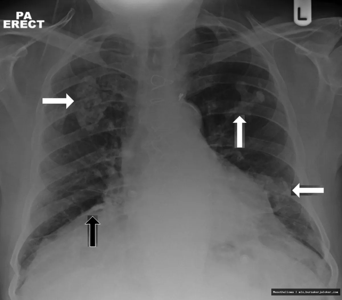
Several abnormalities on a chest X-ray can raise suspicion for mesothelioma. These include:
- Pleural Thickening: This is one of the most common findings. The pleura, the lining around the lungs, appears abnormally thick, often along the chest wall or diaphragm. This thickening can be diffuse (spread out) or localized (in a specific area).
- Pleural Effusions: Fluid accumulation in the pleural space is another frequent finding. The fluid can obscure the underlying lung tissue and make it difficult to visualize other abnormalities.
- Lung Mass or Nodules: While less common, mesothelioma can sometimes present as a mass or nodules within the lung tissue. These may be difficult to distinguish from other lung cancers on an X-ray alone.
- Diaphragmatic Abnormality: Mesothelioma can affect the diaphragm, the muscle that separates the chest and abdomen. X-rays may show elevation or thickening of the diaphragm.
- Asbestos-Related Changes: In some cases, X-rays may reveal signs of asbestos exposure, such as pleural plaques (calcified areas on the pleura) or interstitial lung disease (scarring of the lung tissue). These findings, while not diagnostic of mesothelioma, can increase the suspicion for asbestos-related diseases.
Limitations of X-Rays in Mesothelioma Diagnosis
While X-rays are a valuable initial screening tool, they have significant limitations in diagnosing mesothelioma. These include:
- Low Sensitivity: X-rays are not always sensitive enough to detect early-stage mesothelioma or subtle abnormalities. Small tumors or early pleural thickening may be missed.
- Lack of Specificity: The findings seen on X-rays in mesothelioma can also be caused by other conditions, such as pneumonia, lung cancer, or benign pleural diseases. This makes it difficult to differentiate mesothelioma from other diseases based on X-ray findings alone.
- Limited Soft Tissue Visualization: X-rays are better at visualizing bones than soft tissues. This can make it challenging to assess the extent of tumor involvement or to differentiate between different types of pleural abnormalities.
Due to these limitations, X-rays are typically used as a screening tool to identify patients who require further investigation with more advanced imaging techniques like CT scans or MRIs. A definitive diagnosis of mesothelioma requires a tissue biopsy.
Mesothelioma X-Ray in 2025: Anticipated Advancements
Looking ahead to 2025, several advancements in X-ray technology and image analysis are expected to improve the detection and diagnosis of mesothelioma. These advancements include improvements in image resolution, the integration of artificial intelligence (AI), and the use of contrast agents.
Enhanced Image Resolution and Digital Processing
One of the key areas of improvement in X-ray technology is in image resolution. Newer digital X-ray systems are capable of producing images with greater detail and clarity. This allows radiologists to visualize subtle abnormalities that might have been missed on older systems. Furthermore, advanced digital processing techniques can be used to enhance the contrast and sharpness of X-ray images, making it easier to identify pleural thickening, fluid collections, and other signs of mesothelioma.
Artificial Intelligence (AI) in Mesothelioma Detection
Artificial intelligence (AI) is poised to revolutionize medical imaging, including X-rays. By 2025, AI algorithms are expected to play a significant role in the detection and diagnosis of mesothelioma. These algorithms can be trained to identify subtle patterns and abnormalities on X-rays that may be missed by human radiologists. AI can also help to differentiate between mesothelioma and other conditions that cause similar findings on X-rays.
Specifically, AI can be used to:
- Automated detection of pleural thickening and effusions: AI algorithms can be trained to automatically identify and measure pleural thickening and effusions, providing radiologists with a more objective and consistent assessment of these findings.
- Risk stratification: AI can be used to analyze X-ray images and other clinical data to assess a patient’s risk of developing mesothelioma. This can help to identify individuals who may benefit from earlier screening and intervention.
- Image enhancement and noise reduction: AI algorithms can be used to improve the quality of X-ray images by reducing noise and enhancing contrast. This can make it easier to visualize subtle abnormalities.
Contrast-Enhanced X-Rays
While contrast agents are more commonly used in CT scans and MRIs, there is growing interest in developing contrast agents that can be used with X-rays to improve the visualization of tumors and pleural abnormalities. These contrast agents could help to differentiate between benign and malignant pleural thickening and to assess the extent of tumor involvement. The development of safe and effective contrast agents for X-rays could significantly enhance their diagnostic capabilities in mesothelioma.
The Role of X-Rays in the Mesothelioma Diagnostic Pathway in 2025
Even with these advancements, it’s important to remember that X-rays will likely remain a part of a broader diagnostic pathway. The role of X-rays in mesothelioma diagnosis in 2025 will likely be as follows:
- Initial Screening: X-rays will continue to be used as an initial screening tool for patients with symptoms suggestive of mesothelioma.
- Identifying Suspicious Findings: X-rays will help to identify patients who require further investigation with more advanced imaging techniques.
- Guiding Further Evaluation: X-ray findings can help to guide the selection of appropriate imaging modalities, such as CT scans or MRIs, and to plan for biopsies.
- Monitoring Disease Progression: X-rays may be used to monitor the progression of mesothelioma over time, although CT scans and MRIs are typically preferred for this purpose.
It is crucial to reiterate that a definitive diagnosis of mesothelioma still requires a tissue biopsy. X-rays and other imaging techniques can only suggest the possibility of mesothelioma. A biopsy is necessary to confirm the diagnosis and to determine the type of mesothelioma.
Complementary Imaging Techniques
Given the limitations of X-rays, several complementary imaging techniques are used in the diagnosis and management of mesothelioma. These include:
- CT Scan (Computed Tomography): CT scans provide detailed cross-sectional images of the chest and abdomen, allowing for a more comprehensive assessment of tumor extent and involvement of surrounding structures. CT scans are often used to guide biopsies and to monitor treatment response.
- MRI (Magnetic Resonance Imaging): MRI provides excellent soft tissue contrast and is particularly useful for assessing the involvement of the chest wall, diaphragm, and heart. MRI can also be used to differentiate between different types of pleural abnormalities.
- PET/CT Scan (Positron Emission Tomography/Computed Tomography): PET/CT scans combine the anatomical information from a CT scan with the metabolic information from a PET scan. This can help to identify areas of active tumor growth and to assess treatment response.
These imaging techniques, in conjunction with a tissue biopsy, are essential for making an accurate diagnosis of mesothelioma and for developing an appropriate treatment plan.
Conclusion
While X-rays have limitations in the definitive diagnosis of mesothelioma, they remain a valuable and accessible tool in the initial evaluation process. As we move towards 2025, advancements in X-ray technology, including enhanced image resolution, the integration of artificial intelligence, and the development of contrast agents, are expected to improve their ability to detect suspicious findings and guide further investigation. However, it is important to remember that X-rays are just one piece of the puzzle, and a definitive diagnosis of mesothelioma still requires a tissue biopsy and a multidisciplinary approach involving radiologists, pulmonologists, and oncologists. By understanding the role of X-rays in the mesothelioma diagnostic pathway and by utilizing complementary imaging techniques, healthcare professionals can improve the early detection and management of this challenging disease.
Conclusion
In summary, while mesothelioma X-rays are not definitive diagnostic tools, they play a vital role in the initial stages of detection and monitoring of this aggressive cancer. They can reveal key indicators like pleural thickening, effusions, and masses, prompting further investigation with more specific imaging techniques and biopsies. Understanding the limitations and benefits of X-rays in the context of mesothelioma diagnosis is crucial for both patients and healthcare professionals. The information gleaned from these images, coupled with patient history and other clinical findings, significantly contributes to the overall diagnostic process.
The journey towards diagnosing and managing mesothelioma can be complex and challenging. If you are experiencing symptoms or have a history of asbestos exposure, it is imperative to consult with a medical professional. Early detection is key to improving treatment outcomes. If you have been diagnosed with mesothelioma and require further information or support, resources are available. Please visit the Mesothelioma Applied Research Foundation website for comprehensive information and support services. Remember, you are not alone in this fight.
Frequently Asked Questions (FAQ) about mesothelioma x ray
Can a standard chest x-ray detect mesothelioma, and what are the limitations of using an x-ray for mesothelioma diagnosis?
A standard chest x-ray can sometimes detect abnormalities suggestive of mesothelioma, but it’s generally not considered a definitive diagnostic tool. While an x-ray can reveal pleural thickening, fluid buildup (pleural effusions), or masses in the chest cavity, these findings are not specific to mesothelioma and can be caused by other conditions. Therefore, an x-ray alone is insufficient for a mesothelioma diagnosis. The limitations of using an x-ray include its inability to visualize small tumors or distinguish between mesothelioma and other lung diseases. Furthermore, it may not detect early-stage mesothelioma. If an x-ray suggests mesothelioma, further, more detailed imaging, such as a CT scan or MRI, and ultimately a biopsy, are necessary to confirm the diagnosis.
What specific signs of mesothelioma might a doctor look for on a chest x-ray, and how do these signs differ from those of other lung conditions?
On a chest x-ray, doctors might look for several signs suggestive of mesothelioma. These include pleural thickening (a thickening of the lining of the lung), pleural effusions (fluid buildup between the lung and chest wall), and masses or tumors within the chest cavity. Diffuse pleural thickening, where the thickening is widespread, is more suggestive of mesothelioma than localized thickening. However, these signs can also be present in other lung conditions. For example, pleural thickening can be caused by infections, asbestos exposure without mesothelioma, or other inflammatory conditions. Pleural effusions are also common in pneumonia, heart failure, and other diseases. The key difference lies in the pattern and extent of these findings. Mesothelioma often presents with circumferential pleural thickening and may encase the lung. Because these signs are not unique, further investigation, including CT scans, MRIs, and biopsies, are essential to differentiate mesothelioma from other lung conditions.
If I have been exposed to asbestos, how often should I get a chest x-ray to screen for mesothelioma, and what follow-up tests are recommended if abnormalities are found?
There is no universally agreed-upon guideline for routine chest x-ray screening for mesothelioma in individuals with a history of asbestos exposure. Current medical guidelines generally do not recommend routine x-ray screening due to its limited sensitivity in detecting early-stage mesothelioma and the potential risks associated with radiation exposure. Many doctors instead recommend regular clinical monitoring, which includes discussing any new or concerning symptoms with your doctor. If you experience symptoms like shortness of breath, chest pain, or persistent cough, you should seek medical attention promptly. If an x-ray is performed and abnormalities are found, further diagnostic testing is typically recommended. This may include a CT scan of the chest, which provides more detailed images, or an MRI. Ultimately, a biopsy is necessary to confirm or rule out a diagnosis of mesothelioma. Talk to your doctor about the best monitoring plan for you based on your individual risk factors and medical history.