Mesothelioma USMLE: 2025 Prep & Key Exam Points
The USMLE (United States Medical Licensing Examination) is a critical hurdle for medical students and graduates aiming to practice medicine in the United States. While the USMLE covers a vast range of medical topics, certain diseases, like mesothelioma, often appear due to their clinical significance and potential for complex presentations. Understanding the key aspects of mesothelioma, its pathogenesis, diagnosis, and management, is crucial for success on the USMLE. This article provides a comprehensive overview of mesothelioma as it relates to USMLE preparation, focusing on high-yield information and key exam points relevant for the 2025 USMLE exams.
Mesothelioma, a rare and aggressive cancer arising from the mesothelial cells lining the pleura, peritoneum, pericardium, and tunica vaginalis, is strongly associated with asbestos exposure. Its insidious onset and often vague initial symptoms make early diagnosis challenging, contributing to its poor prognosis. For the USMLE, you’ll need to grasp the disease’s epidemiology, risk factors, pathophysiology, clinical presentation, diagnostic approaches, and treatment strategies. This includes recognizing the different histological subtypes and understanding the genetic mutations that contribute to its development.
This article will guide you through the essential information about mesothelioma that you’ll need to know for the USMLE, specifically tailored for the 2025 exam. We will delve into the key concepts, diagnostic modalities, and treatment options that are frequently tested. By focusing on the most relevant aspects and providing practical tips, this guide aims to equip you with the knowledge and confidence to tackle mesothelioma-related questions effectively and improve your overall USMLE performance. So, let’s begin our exploration of mesothelioma in the context of the USMLE.
Mesothelioma USMLE: Epidemiology and Risk Factors
Understanding the epidemiology and risk factors of mesothelioma is crucial for answering USMLE questions that test your ability to identify patients at risk and interpret clinical scenarios. Asbestos exposure is, by far, the most significant risk factor. However, it’s important to remember that not everyone exposed to asbestos will develop mesothelioma, and other factors can play a role.
Asbestos Exposure: The Primary Culprit
Asbestos, a naturally occurring mineral fiber, was widely used in various industries for its heat resistance and insulation properties. Inhalation of asbestos fibers leads to chronic inflammation and DNA damage in mesothelial cells, eventually leading to malignant transformation. Occupations at high risk of asbestos exposure include:
- Construction workers
- Shipyard workers
- Insulation installers
- Miners
- Automotive mechanics (brake linings)
The latency period between asbestos exposure and the development of mesothelioma can be very long, often spanning 20-50 years. This long latency period makes it challenging to trace the source of exposure in some cases.
Other Risk Factors and Considerations
While asbestos is the dominant risk factor, other factors can contribute to the development of mesothelioma, although they are much less common:
- Eruonite: A fibrous mineral similar to asbestos, found in certain parts of the world.
- Radiation exposure: High doses of radiation, particularly to the chest, have been linked to an increased risk.
- SV40 virus: Some studies have suggested a possible link between the simian virus 40 (SV40) and mesothelioma, although this remains controversial.
- Genetic predisposition: Rare cases of familial mesothelioma have been reported, suggesting a possible genetic susceptibility in some individuals, particularly mutations in the BAP1 gene.
USMLE Tip: Be prepared to identify patients with a history of asbestos exposure based on their occupation or residence. Remember the long latency period when evaluating a patient with potential mesothelioma symptoms.
Mesothelioma USMLE: Pathophysiology and Histopathology
A strong understanding of the underlying mechanisms and histological subtypes of mesothelioma is essential for USMLE success. This section will cover the key pathophysiological processes and the different histological variants, which are frequently tested.
Pathophysiological Mechanisms
Asbestos fibers, once inhaled, reach the pleura or peritoneum and are phagocytosed by macrophages. This process leads to chronic inflammation, oxidative stress, and the release of growth factors that promote mesothelial cell proliferation. Key molecular pathways involved in mesothelioma development include:
- BAP1 inactivation: Mutations in the BAP1 gene are frequently found in mesothelioma. BAP1 is a tumor suppressor gene involved in DNA repair and chromatin regulation.
- NF2 inactivation: Mutations in the NF2 gene, which encodes merlin (schwannomin), a tumor suppressor protein, are also common.
- CDKN2A deletion: Deletion of the CDKN2A gene, which encodes p16 and p14ARF, cell cycle regulators, is frequently observed.
- Hippo signaling pathway: Deregulation of the Hippo signaling pathway, which controls cell growth and proliferation, is implicated in mesothelioma pathogenesis.
These genetic alterations disrupt normal cell cycle control, DNA repair mechanisms, and apoptosis, leading to uncontrolled cell growth and tumor formation.
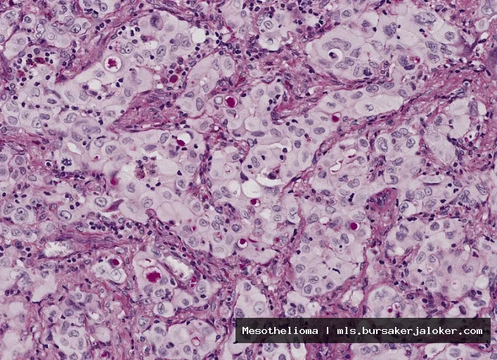
Histological Subtypes of Mesothelioma
Mesothelioma is classified into different histological subtypes, each with distinct microscopic features and prognostic implications. The three main subtypes are:
- Epithelioid: The most common subtype (50-70% of cases). Characterized by cells with epithelial-like features, such as cuboidal or polygonal shape and well-defined cell borders. This subtype generally has a better prognosis compared to the sarcomatoid subtype.
- Sarcomatoid: A less common subtype (10-20% of cases). Composed of spindle-shaped cells that resemble fibroblasts. This subtype is more aggressive and associated with a poorer prognosis.
- Biphasic (Mixed): Contains both epithelioid and sarcomatoid components. The prognosis depends on the proportion of each component, with a higher sarcomatoid component associated with a worse outcome.
USMLE Tip: Be able to differentiate between the histological subtypes based on their microscopic features. Understand that the sarcomatoid subtype has the worst prognosis.
Mesothelioma USMLE: Clinical Presentation, Diagnosis, and Staging
This section focuses on the clinical manifestations, diagnostic procedures, and staging systems used for mesothelioma, all of which are crucial for accurate diagnosis and appropriate management. Mastering these aspects will significantly improve your performance on USMLE questions related to mesothelioma.
Clinical Presentation
The clinical presentation of mesothelioma varies depending on the site of origin. Common symptoms include:
- Pleural Mesothelioma:
- Chest pain
- Shortness of breath (dyspnea)
- Cough
- Pleural effusion
- Weight loss
- Fatigue
- Peritoneal Mesothelioma:
- Abdominal pain
- Abdominal swelling (ascites)
- Bowel obstruction
- Weight loss
Paraneoplastic syndromes, such as thrombocytosis or hypercalcemia, can also occur in some cases.
Diagnostic Procedures
The diagnosis of mesothelioma typically involves a combination of imaging studies, cytology, and tissue biopsy. Key diagnostic procedures include:
- Imaging Studies:
- Chest X-ray: May show pleural thickening, pleural effusion, or masses.
- CT scan: Provides more detailed images of the chest and abdomen, helping to assess the extent of the disease.
- MRI: Can be useful for evaluating local invasion and distinguishing mesothelioma from other pleural diseases.
- PET/CT scan: Can help assess the metabolic activity of the tumor and identify metastatic disease.
- Cytology:
- Pleural fluid cytology: Analysis of pleural fluid obtained by thoracentesis can sometimes reveal malignant mesothelial cells, but the sensitivity is relatively low.
- Peritoneal fluid cytology: Analysis of ascitic fluid obtained by paracentesis can also detect malignant mesothelial cells.
- Tissue Biopsy:
- Thoracoscopy/Laparoscopy: A minimally invasive procedure to obtain tissue samples from the pleura or peritoneum.
- Surgical biopsy: Open surgical biopsy may be necessary to obtain sufficient tissue for diagnosis, especially if thoracoscopy or laparoscopy is not feasible.
Immunohistochemical staining is crucial for confirming the diagnosis of mesothelioma and differentiating it from other malignancies. Common markers used include calretinin, cytokeratin 5/6, WT1, and D2-40.
Staging
The most commonly used staging system for pleural mesothelioma is the TNM (Tumor, Node, Metastasis) staging system. The staging system considers the size and extent of the primary tumor (T), the presence or absence of lymph node involvement (N), and the presence or absence of distant metastasis (M). Staging is important for determining prognosis and guiding treatment decisions.
USMLE Tip: Understand the common presenting symptoms of pleural and peritoneal mesothelioma. Know the role of imaging studies, cytology, and tissue biopsy in the diagnostic process. Be familiar with the TNM staging system for pleural mesothelioma.
Mesothelioma USMLE: Treatment and Management
This section will cover the different treatment modalities used for mesothelioma and their respective roles in managing the disease. Understanding the principles of treatment and the factors that influence treatment decisions is essential for answering USMLE questions related to management strategies.
Treatment Modalities
The treatment of mesothelioma is often multimodal, involving a combination of surgery, chemotherapy, and radiation therapy. The specific treatment approach depends on the stage of the disease, the patient’s overall health, and the histological subtype of the tumor.
- Surgery:
- Extrapleural pneumonectomy (EPP): Involves the removal of the entire lung, pleura, pericardium, and diaphragm on the affected side. This is a radical surgery and is typically reserved for patients with early-stage disease who are otherwise healthy.
- Pleurectomy/decortication (P/D): Involves the removal of the pleura while preserving the lung. This is a less radical surgery than EPP and may be suitable for patients who are not candidates for EPP.
- Chemotherapy:
- Pemetrexed and cisplatin: The standard first-line chemotherapy regimen for mesothelioma.
- Pemetrexed and carboplatin: An alternative regimen for patients who cannot tolerate cisplatin.
- Radiation Therapy:
Can be used to control local tumor growth and relieve symptoms. It is often used as an adjunct to surgery or chemotherapy.
- Immunotherapy:
Immune checkpoint inhibitors, such as nivolumab and ipilimumab, have shown promising results in treating mesothelioma, particularly in patients who have progressed on chemotherapy.
Factors Influencing Treatment Decisions
Several factors influence treatment decisions in mesothelioma, including:
- Stage of the disease: Early-stage disease is more likely to be amenable to surgical resection.
- Histological subtype: Epithelioid mesothelioma generally responds better to treatment than sarcomatoid mesothelioma.
- Patient’s overall health: Patients with good performance status are more likely to tolerate aggressive treatment.
- Presence of comorbidities: Underlying medical conditions can affect treatment options.
Palliative Care
Palliative care plays a crucial role in managing symptoms and improving the quality of life for patients with mesothelioma. Palliative measures include pain management, symptom control, and emotional support.
USMLE Tip: Know the different treatment modalities used for mesothelioma and their indications. Understand the role of palliative care in managing symptoms and improving quality of life. Be aware of the common chemotherapy regimens used for mesothelioma.
Mesothelioma USMLE: Key Takeaways and Practice Questions
This final section summarizes the key points discussed in this article and provides practice questions to help you assess your understanding of mesothelioma in the context of the USMLE.
Key Takeaways
- Asbestos exposure is the primary risk factor for mesothelioma.
- The latency period between asbestos exposure and mesothelioma development can be very long.
- Mesothelioma can arise in the pleura, peritoneum, pericardium, and tunica vaginalis.
- The three main histological subtypes of mesothelioma are epithelioid, sarcomatoid, and biphasic.
- The sarcomatoid subtype has the worst prognosis.
- Diagnosis involves imaging studies, cytology, and tissue biopsy.
- Treatment is often multimodal, involving surgery, chemotherapy, and radiation therapy.
- Pemetrexed and cisplatin are the standard first-line chemotherapy regimen.
Practice Questions
- A 65-year-old man presents with chest pain and shortness of breath. He reports a history of working as a shipyard worker for 30 years. Chest X-ray reveals pleural thickening and a large pleural effusion. What is the most likely diagnosis?
- Pneumonia
- Lung cancer
- Mesothelioma
- Tuberculosis
- Which of the following is the most common histological subtype of mesothelioma?
- Sarcomatoid
- Epithelioid
- Biphasic
- Desmoplastic
- A patient with mesothelioma is being treated with pemetrexed and cisplatin. What is the most common side effect of cisplatin?
- Alopecia
- Nephrotoxicity
- Cardiotoxicity
- Pulmonary fibrosis
(Answers: 1. c, 2. b, 3. b). For more information, you can refer to Mesothelioma as an additional resource.
By mastering the information presented in this article and practicing with relevant questions, you will be well-prepared to tackle mesothelioma-related questions on the USMLE and improve your overall exam performance. Good luck with your studies!
Conclusion
In summary, successfully navigating mesothelioma-related questions on the USMLE requires a multifaceted approach. This includes a solid understanding of the disease’s etiology, particularly its strong association with asbestos exposure, its clinical presentation encompassing symptoms like dyspnea and chest pain, and the various diagnostic modalities employed, such as imaging techniques and tissue biopsies. Furthermore, familiarity with the different histological subtypes and their prognostic implications is crucial. Mastering these key areas will significantly enhance your performance on USMLE exams and, more importantly, prepare you for the challenges of diagnosing and managing patients with this complex malignancy in clinical practice.
Ultimately, a dedicated and comprehensive study strategy focusing on these critical aspects of mesothelioma is essential for achieving success on the USMLE. We encourage you to utilize the resources mentioned throughout this article, consult with experienced mentors, and engage in thorough practice questions. By doing so, you will not only improve your exam score but also develop a deeper understanding of this devastating disease, allowing you to provide better care for future patients. For further information and updated guidelines on mesothelioma diagnosis and treatment, please refer to reputable medical organizations such as the American Cancer Society.
Frequently Asked Questions (FAQ) about mesothelioma usmle
How might a question about mesothelioma present on the USMLE Step 1 exam, and what key histological findings should I look for to distinguish it from other cancers?
Mesothelioma on the USMLE Step 1 is often presented as a clinical vignette involving a patient with a history of asbestos exposure, presenting with symptoms like shortness of breath, chest pain, and pleural effusions. The question may ask about the most likely diagnosis or the underlying pathophysiology. Key histological findings are crucial for differentiation. Look for biphasic or epithelioid and sarcomatoid patterns. Calretinin and cytokeratin 5/6 are typically positive immunohistochemical markers, while Ber-EP4 is negative, helping to distinguish it from adenocarcinoma. Understanding the association with asbestos exposure and recognizing these specific histological and immunohistochemical features are essential for answering such questions correctly.
What are the most important risk factors for developing mesothelioma that I should remember for the USMLE Step exams, and how does asbestos exposure specifically contribute to the pathogenesis of this cancer?
The most important risk factor for mesothelioma, crucial for the USMLE Step exams, is overwhelmingly asbestos exposure. While other factors like simian virus 40 (SV40) have been investigated, asbestos remains the primary and most well-established cause. Asbestos fibers, when inhaled, lodge in the pleura or peritoneum, triggering chronic inflammation and DNA damage. This chronic inflammation leads to the release of reactive oxygen species and other inflammatory mediators, promoting cellular proliferation and ultimately leading to the development of mesothelioma. The latency period between asbestos exposure and the development of mesothelioma can be very long, often decades. Therefore, a history of asbestos exposure, even in the distant past, is a critical clue in the diagnosis.
Beyond histology, what imaging modalities and laboratory tests are commonly used in the diagnosis of mesothelioma, and how might these be presented in a USMLE Step 2 CK or Step 3 question?
Besides histology, diagnostic workup for mesothelioma involves imaging and laboratory tests. Chest X-rays and CT scans are commonly used to identify pleural thickening, pleural effusions, and masses. MRI can provide more detailed information about the extent of the disease. Pleural fluid analysis, obtained via thoracentesis, may reveal malignant cells, but is often non-diagnostic. Soluble mesothelin-related protein (SMRP) is a serum marker that can be elevated in mesothelioma, though it’s not highly sensitive or specific. On USMLE Step 2 CK or Step 3, you might see a vignette describing a patient with pleural effusion. The question could ask about the next best step in diagnosis, which would likely be a CT scan of the chest followed by a biopsy of the pleura for histological confirmation. SMRP might be mentioned as a supportive, but not definitive, diagnostic tool. Be sure to understand the role and limitations of each diagnostic modality.