Mesothelioma X-Ray Findings: What to Look For (2025)
Mesothelioma, a rare and aggressive cancer primarily affecting the lining of the lungs, abdomen, or heart, often presents diagnostic challenges. While advanced imaging techniques like CT scans and MRIs are crucial for definitive diagnosis and staging, chest X-rays remain a valuable initial screening tool. In 2025, with advancements in digital radiography and image interpretation, understanding the subtle yet significant X-ray findings associated with mesothelioma is more important than ever for early detection and improved patient outcomes. This article will delve into the key radiographic features that clinicians look for when evaluating a chest X-ray for potential mesothelioma.
It’s important to remember that X-rays are often the first imaging study performed when a patient presents with symptoms like shortness of breath, chest pain, or persistent cough. While an X-ray alone cannot definitively diagnose mesothelioma, it can raise suspicion and prompt further investigation with more specialized imaging and biopsies. Recognizing the patterns and abnormalities on an X-ray that are commonly associated with mesothelioma is crucial for timely referral to a specialist and initiation of appropriate diagnostic procedures. This article aims to provide a comprehensive overview of these findings, enabling healthcare professionals to better interpret chest X-rays and contribute to the early detection of this devastating disease.
This article will not only discuss the common radiographic findings but also explore the limitations of X-rays in diagnosing mesothelioma. We will consider how mesothelioma can mimic other conditions on X-ray, such as pleural effusions caused by pneumonia or congestive heart failure. Furthermore, we will highlight the importance of considering the patient’s history, particularly any known asbestos exposure, in conjunction with the radiographic findings. By understanding the nuances of mesothelioma X-ray findings, clinicians can make more informed decisions regarding further diagnostic workup and ultimately improve patient care.
Mesothelioma X-Ray Findings: What to Look For (2025)
Chest X-rays are a fundamental imaging modality used to evaluate the lungs and surrounding structures. In the context of mesothelioma, several characteristic findings can raise suspicion and warrant further investigation. However, it’s crucial to remember that these findings can be subtle and may overlap with other conditions, necessitating a thorough clinical evaluation and correlation with other diagnostic tests.
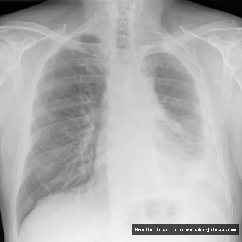
Pleural Thickening
One of the most common and significant X-ray findings in mesothelioma is pleural thickening. The pleura is the thin membrane that lines the lungs and the inside of the chest wall. Mesothelioma often originates in the pleura, causing it to thicken and become irregular. On an X-ray, this thickening appears as a hazy or dense area along the lung periphery. It’s important to distinguish pleural thickening from normal anatomical structures or other causes of pleural disease, such as infection or inflammation.
- Circumferential Pleural Thickening: This refers to thickening that extends around a significant portion of the lung. It’s a strong indicator of mesothelioma, particularly when accompanied by other findings.
- Nodular Pleural Thickening: Irregular, bumpy thickening is more suggestive of malignancy than smooth, uniform thickening. These nodules can be difficult to visualize on X-ray alone and may require CT scanning for better characterization.
- Pleural Plaques: While pleural plaques themselves are not diagnostic of mesothelioma, their presence can indicate asbestos exposure, which is a major risk factor for the disease. Plaques appear as calcified or non-calcified areas of thickening along the pleura.
Pleural Effusion
Pleural effusion, the accumulation of fluid in the pleural space, is another common finding in mesothelioma. The fluid can compress the lung and cause shortness of breath. On an X-ray, a pleural effusion appears as a homogenous density that obscures the underlying lung tissue. The presence of a large effusion can make it difficult to visualize other underlying abnormalities, such as pleural thickening or masses.
- Loculated Effusion: Unlike a free-flowing effusion, a loculated effusion is trapped in pockets within the pleural space. This can be caused by adhesions or scarring from previous inflammation or infection. Loculated effusions are more commonly associated with mesothelioma.
- Effusion Recurrence: Mesothelioma-related effusions often recur despite drainage, a characteristic that can help differentiate them from effusions caused by other conditions.
Lung Volume Loss
As mesothelioma progresses, it can restrict lung expansion and lead to lung volume loss. This can be seen on an X-ray as a smaller lung size on the affected side compared to the unaffected side. The mediastinum (the space in the chest containing the heart and major blood vessels) may also shift towards the affected side.
Mediastinal Involvement
In some cases, mesothelioma can spread to the mediastinum, the space between the lungs. This can manifest on an X-ray as widening of the mediastinum or the presence of mediastinal masses. Mediastinal involvement is a sign of more advanced disease.
Diaphragmatic Abnormality
Mesothelioma can affect the diaphragm, the muscle that separates the chest from the abdomen. This can lead to elevation or irregularity of the diaphragm on an X-ray. In advanced cases, the tumor can even invade the diaphragm.
Limitations of X-Rays in Diagnosing Mesothelioma
While chest X-rays are a valuable initial screening tool, they have limitations in diagnosing mesothelioma. Several factors can make it challenging to accurately detect and characterize the disease on X-ray alone.
Sensitivity and Specificity
X-rays have relatively low sensitivity and specificity for detecting early mesothelioma. Small pleural abnormalities can be easily missed, and the findings can be nonspecific, mimicking other conditions. A high degree of suspicion is required, particularly in patients with a history of asbestos exposure.
Overlapping Conditions
Many other conditions can cause similar findings on chest X-ray, such as pleural effusions from pneumonia, congestive heart failure, or other malignancies. Pleural thickening can also be caused by benign conditions like asbestos-related pleural plaques or post-inflammatory changes. Differentiating mesothelioma from these conditions requires careful clinical evaluation and often necessitates further imaging studies.
Lesion Location and Size
Small or peripherally located lesions may be difficult to visualize on X-ray. Similarly, lesions hidden behind the heart or diaphragm can be obscured. Larger lesions are more easily detected, but by this stage, the disease is often more advanced.
Image Quality and Interpretation
The quality of the X-ray image and the experience of the radiologist interpreting the image can also affect the accuracy of the diagnosis. Poor image quality can make it difficult to visualize subtle abnormalities, and inexperienced radiologists may miss early signs of mesothelioma.
The Role of X-Rays in Conjunction with Other Diagnostic Tools
Given the limitations of X-rays in diagnosing mesothelioma, it’s crucial to use them in conjunction with other diagnostic tools to confirm the diagnosis and determine the extent of the disease. The following are some of the key diagnostic modalities used in the evaluation of suspected mesothelioma:
CT Scans
Computed tomography (CT) scans are more sensitive and specific than X-rays for detecting and characterizing pleural abnormalities. CT scans can provide detailed images of the chest, including the pleura, lungs, mediastinum, and diaphragm. They can also help to differentiate mesothelioma from other conditions and to assess the extent of the disease. Understanding the complexities of asbestos exposure is crucial, Mesothelioma a rare and aggressive cancer affecting the lining of the lungs, abdomen, or heart
.
MRI Scans
Magnetic resonance imaging (MRI) scans can provide even more detailed images of the soft tissues in the chest. MRI is particularly useful for evaluating the chest wall invasion and diaphragmatic involvement. It can also help to differentiate mesothelioma from other types of tumors.
PET Scans
Positron emission tomography (PET) scans can help to detect metabolically active cancer cells. PET scans are often used in conjunction with CT scans (PET/CT) to assess the extent of the disease and to monitor the response to treatment.
Biopsy
The only way to definitively diagnose mesothelioma is through a biopsy. A biopsy involves taking a small sample of tissue from the pleura and examining it under a microscope. There are several different types of biopsies that can be used to diagnose mesothelioma, including:
- Thoracentesis: Removing fluid from the pleural space for analysis.
- Pleural Biopsy: Taking a small sample of the pleura using a needle or during surgery.
- Thoracoscopy: A minimally invasive procedure that allows the surgeon to visualize the pleura and take biopsies.
- Open Lung Biopsy: A more invasive procedure that involves surgically opening the chest to take a biopsy.
Conclusion: Optimizing Mesothelioma Detection in 2025
In 2025, while advanced imaging technologies are readily available, chest X-rays remain a vital first-line investigation for patients presenting with respiratory symptoms. Understanding the characteristic X-ray findings associated with mesothelioma – pleural thickening, pleural effusion, lung volume loss, mediastinal involvement, and diaphragmatic abnormalities – is crucial for early detection. However, it is equally important to recognize the limitations of X-rays and to use them in conjunction with other diagnostic tools, such as CT scans, MRI scans, PET scans, and biopsies, to confirm the diagnosis and determine the extent of the disease. Considering the patient’s history, particularly any asbestos exposure, is also essential for accurate interpretation of radiographic findings.
By staying abreast of the latest advancements in imaging technology and diagnostic techniques, healthcare professionals can improve their ability to detect mesothelioma early, leading to more effective treatment and improved patient outcomes. Continued research and education are essential to further refine our understanding of mesothelioma and to develop new and improved methods for diagnosis and treatment.
Ultimately, a multidisciplinary approach involving radiologists, pulmonologists, oncologists, and surgeons is crucial for providing the best possible care for patients with mesothelioma. Early detection and accurate diagnosis are key to improving the prognosis for this devastating disease. By leveraging the power of X-rays in conjunction with other diagnostic modalities, we can strive to achieve earlier and more accurate diagnoses, ultimately improving the lives of patients affected by mesothelioma.
Conclusion
In conclusion, while chest X-rays play a crucial role in the initial assessment of patients suspected of having mesothelioma, their limitations must be acknowledged. Although characteristic findings such as pleural thickening, pleural effusions, and potential lung volume loss can raise suspicion, they are not definitive for diagnosis. The presence of these radiographic features necessitates further investigation with more advanced imaging techniques like CT scans and ultimately, tissue biopsy for confirmation. Therefore, relying solely on X-ray findings can lead to both delayed diagnosis and misdiagnosis, highlighting the importance of a comprehensive diagnostic approach.
The information presented here emphasizes the significance of understanding the role of X-rays in the diagnostic pathway for mesothelioma. If you are experiencing symptoms such as shortness of breath, chest pain, or persistent cough, especially with a history of asbestos exposure, it is crucial to consult with a qualified medical professional. Early detection and accurate diagnosis are paramount for effective treatment and improved outcomes. Learn more about mesothelioma diagnosis and treatment options by visiting the Mesothelioma Applied Research Foundation website.
Frequently Asked Questions (FAQ) about mesothelioma x ray findings
What are the common mesothelioma x-ray findings that doctors look for when diagnosing this cancer?
When evaluating a chest x-ray for possible mesothelioma, doctors look for several characteristic findings. One of the most common is pleural thickening, which appears as an abnormal density along the lining of the lung (pleura). This thickening can be diffuse (spread out) or nodular (lumpy). Another key finding is pleural effusion, which is an accumulation of fluid in the pleural space. The x-ray may also reveal a shrunken lung volume on the affected side. In advanced cases, the x-ray might show signs of the tumor invading nearby structures, such as the diaphragm or chest wall. It’s important to note that x-ray findings alone are not enough to diagnose mesothelioma; further imaging, like CT scans or MRI, and a biopsy are necessary for definitive diagnosis.
How accurate are x-rays in detecting mesothelioma, and what are the limitations of using x-rays to diagnose this disease?
While chest x-rays are often the first imaging test used when mesothelioma is suspected, they have limitations in accuracy for diagnosing the disease. X-rays are good at detecting pleural effusions (fluid around the lungs) and significant pleural thickening, which are common signs of mesothelioma. However, they may miss smaller tumors or subtle changes in the pleura. Furthermore, other conditions can cause similar x-ray findings, leading to false positives. Therefore, x-rays are typically used as an initial screening tool. Due to their limitations, additional imaging techniques like CT scans and MRI are crucial for a more detailed assessment. A biopsy is ultimately required for a definitive mesothelioma diagnosis. The sensitivity of x-rays is lower compared to more advanced imaging, so relying solely on x-rays can delay diagnosis.
If I have a normal chest x-ray, does that mean I definitely don’t have mesothelioma, especially if I have a history of asbestos exposure?
A normal chest x-ray does not definitively rule out mesothelioma, particularly if you have a history of asbestos exposure. While x-rays can detect some signs of mesothelioma, such as pleural thickening or effusions, they are not sensitive enough to identify all cases, especially in the early stages. Mesothelioma can sometimes present with subtle changes that are not easily visible on an x-ray. Also, other conditions can mask the presence of a tumor. Given your asbestos exposure history, it’s crucial to discuss your concerns with your doctor. They may recommend more sensitive imaging tests, such as a CT scan or MRI, even if your x-ray is normal, to provide a more thorough evaluation and to monitor for any potential development of asbestos-related diseases, including mesothelioma. Early detection is critical for better treatment outcomes.