Mesothelioma Gross: What You Need to Know
The term “mesothelioma gross” might sound jarring, even insensitive. It’s often used to describe the macroscopic appearance of mesothelioma tumors and the effects this cancer has on the body’s tissues and organs. While it’s crucial to approach this topic with sensitivity and respect for those affected, understanding the physical manifestations of mesothelioma is essential for medical professionals, researchers, and even those seeking information about the disease for themselves or loved ones. This article aims to provide a factual and informative overview of the macroscopic features of mesothelioma, focusing on its appearance, growth patterns, and impact on the body, while maintaining a compassionate and respectful tone.
Mesothelioma is a rare and aggressive cancer that develops in the mesothelium, a protective lining that covers most of the body’s internal organs. The most common type, pleural mesothelioma, affects the lining of the lungs. Peritoneal mesothelioma affects the lining of the abdomen, while pericardial mesothelioma affects the lining of the heart, and tunica vaginalis mesothelioma affects the lining of the testicles. Each type presents with its own unique set of macroscopic characteristics, influenced by the location and extent of the tumor’s growth. Understanding these characteristics helps in diagnosis, staging, and treatment planning.
This article will delve into the various aspects of “mesothelioma gross,” including the appearance of tumors during surgery or autopsy, the changes in affected organs, and the overall impact on the body. It’s important to remember that visualizing and understanding these aspects of the disease, while potentially disturbing, is vital for advancing medical knowledge and improving patient outcomes. We will explore the textures, colors, and growth patterns associated with mesothelioma, providing a comprehensive overview of the macroscopic features of this devastating disease. We aim to provide accurate information while acknowledging the sensitive nature of the topic.
Understanding Mesothelioma: A Macroscopic Perspective
When discussing the “gross” appearance of mesothelioma, we are referring to what can be observed with the naked eye during surgical procedures, autopsies, or through imaging techniques like CT scans and MRIs. This macroscopic view is crucial for pathologists and surgeons as they assess the extent of the disease, determine the stage of cancer, and plan treatment strategies. The appearance can vary greatly depending on the type of mesothelioma, the stage of the disease, and the individual patient. It’s important to remember that these are medical observations, and the term “gross” is used in a clinical context, not to diminish the suffering of those affected.
Pleural Mesothelioma: The Lining of the Lungs
Pleural mesothelioma, affecting the lining of the lungs (pleura), is the most common form of the disease. Macroscopically, it often presents as thick, white or grayish-white plaques or nodules that spread across the pleural surface. These plaques can coalesce and encase the lung, restricting its ability to expand and contract. The tumor can also invade the chest wall, diaphragm, and mediastinum (the space between the lungs). The pleura often appears thickened and fibrotic, losing its normal smooth and glistening appearance. In advanced stages, large, bulky masses may be visible, and fluid accumulation (pleural effusion) is common, further compressing the lung.
Peritoneal Mesothelioma: The Lining of the Abdomen
Peritoneal mesothelioma, affecting the lining of the abdomen (peritoneum), typically presents as numerous small nodules or masses scattered throughout the abdominal cavity. These nodules can range in size from a few millimeters to several centimeters. The peritoneum often appears thickened and covered in a grayish-white or yellowish-white coating. Unlike pleural mesothelioma, which tends to form a solid mass, peritoneal mesothelioma often has a more diffuse and nodular pattern of growth. Ascites, the accumulation of fluid in the abdominal cavity, is a common feature, contributing to abdominal swelling and discomfort. The tumor can also encase abdominal organs, such as the intestines, liver, and spleen.
Pericardial Mesothelioma: The Lining of the Heart
Pericardial mesothelioma, affecting the lining of the heart (pericardium), is a very rare form of the disease. Macroscopically, it often presents as a thickening of the pericardium, with the formation of nodules or masses around the heart. The pericardium may appear fibrotic and constricted, limiting the heart’s ability to pump blood effectively. Pericardial effusion, the accumulation of fluid around the heart, is a common finding, leading to cardiac tamponade, a life-threatening condition in which the heart is compressed. The tumor can also invade the heart muscle itself.
Tunica Vaginalis Mesothelioma: The Lining of the Testicles
Tunica vaginalis mesothelioma, affecting the lining of the testicles (tunica vaginalis), is another rare form of the disease. It typically presents as a mass or swelling within the scrotum. The tunica vaginalis may appear thickened and nodular, and the tumor can invade the testicle itself. Hydrocele, the accumulation of fluid around the testicle, is often present. The macroscopic appearance can sometimes be confused with other testicular masses, making accurate diagnosis crucial.
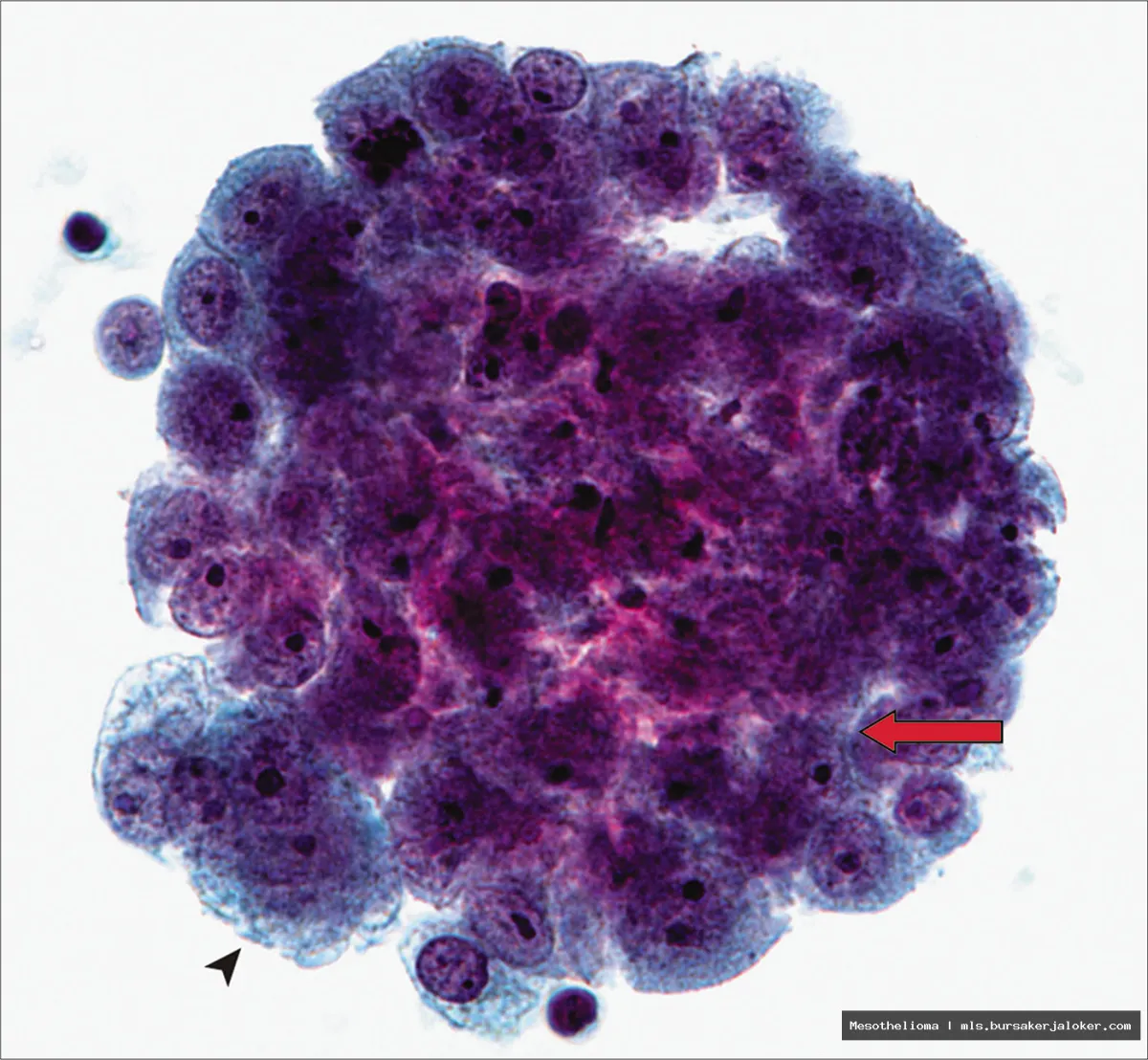
Cellular Level: Microscopic Features of Mesothelioma
While the “gross” appearance refers to what’s visible with the naked eye, examining mesothelioma at the microscopic level is equally important for diagnosis and understanding the disease’s behavior. Histopathology, the microscopic study of tissues, reveals distinct cellular features that help differentiate mesothelioma from other types of cancer. These features are crucial for confirming the diagnosis and determining the specific subtype of mesothelioma.
Epithelioid Mesothelioma
Epithelioid mesothelioma is the most common subtype. Under the microscope, it is characterized by cells that resemble epithelial cells, often arranged in tubular, papillary, or solid patterns. The cells are typically polygonal or cuboidal in shape, with distinct cell borders and relatively abundant cytoplasm. The nuclei are often round or oval, with prominent nucleoli (structures within the nucleus). Immunohistochemical stains, which use antibodies to identify specific proteins in the cells, are essential for confirming the diagnosis and distinguishing epithelioid mesothelioma from other carcinomas.
Sarcomatoid Mesothelioma
Sarcomatoid mesothelioma is a less common and more aggressive subtype. Under the microscope, it is characterized by spindle-shaped cells that resemble fibroblasts or sarcomas. These cells are typically arranged in fascicles (bundles) or sheets, and they often have elongated nuclei. Sarcomatoid mesothelioma can be challenging to diagnose because it can resemble other sarcomas. Immunohistochemical stains are crucial for differentiating it from other spindle cell tumors.
Biphasic Mesothelioma
Biphasic mesothelioma contains both epithelioid and sarcomatoid components. The proportions of each component can vary, and the diagnosis requires the presence of both cell types. Biphasic mesothelioma can be challenging to diagnose because the two components may not be equally represented in all areas of the tumor. Immunohistochemical stains are used to confirm the presence of both epithelioid and sarcomatoid cells.
Impact of Mesothelioma on the Body
The macroscopic growth patterns of mesothelioma directly impact the function of the affected organs and the overall health of the patient. The encasement of organs, the accumulation of fluid, and the invasion of surrounding tissues all contribute to the symptoms and complications associated with the disease. Understanding these impacts is crucial for managing the disease and improving the patient’s quality of life.
Respiratory Complications
Pleural mesothelioma, in particular, significantly impacts respiratory function. The thickening of the pleura and the formation of tumors restrict lung expansion, leading to shortness of breath (dyspnea). Pleural effusion further compresses the lung, exacerbating breathing difficulties. The tumor can also invade the chest wall, causing pain and discomfort. In advanced stages, the tumor can obstruct the airways, leading to pneumonia and other respiratory infections.
Abdominal Complications
Peritoneal mesothelioma causes a range of abdominal complications. Ascites leads to abdominal swelling, pain, and discomfort. The tumor can also encase abdominal organs, leading to bowel obstruction, nausea, vomiting, and malnutrition. The tumor can also compress blood vessels, leading to edema (swelling) in the legs and ankles. In advanced stages, the tumor can invade the liver and other organs, causing organ failure. For more information, you can refer to Mesothelioma as an additional resource.
Cardiac Complications
Pericardial mesothelioma, although rare, can have devastating effects on the heart. Pericardial effusion can lead to cardiac tamponade, a life-threatening condition in which the heart is compressed, preventing it from pumping blood effectively. The tumor can also invade the heart muscle, leading to arrhythmias (irregular heartbeats) and heart failure. The constriction of the pericardium can also limit the heart’s ability to fill with blood, leading to reduced cardiac output.
Diagnosis and Imaging Techniques
While the “gross” appearance of mesothelioma can be observed during surgery or autopsy, imaging techniques play a crucial role in diagnosis and staging. These techniques allow doctors to visualize the tumor and assess its extent before invasive procedures are performed. Common imaging techniques include:
- CT Scans (Computed Tomography): CT scans provide detailed cross-sectional images of the body, allowing doctors to visualize the tumor, assess its size and location, and identify any invasion of surrounding tissues.
- MRIs (Magnetic Resonance Imaging): MRIs use magnetic fields and radio waves to create detailed images of the body. MRIs are particularly useful for visualizing soft tissues and can provide more detailed information about the tumor’s extent and its relationship to surrounding structures.
- PET Scans (Positron Emission Tomography): PET scans use radioactive tracers to detect metabolically active cells. PET scans can help identify areas of cancer that may not be visible on CT or MRI scans.
- X-Rays: Chest X-rays can help detect pleural effusion and other abnormalities in the lungs.
Conclusion
Understanding the “gross” appearance of mesothelioma, while a sensitive topic, is essential for medical professionals, researchers, and those affected by the disease. By understanding the macroscopic features of mesothelioma, we can improve diagnosis, staging, and treatment planning. While the physical manifestations of this cancer can be disturbing, it’s important to remember that this knowledge is crucial for advancing medical knowledge and improving patient outcomes. Approaching this topic with sensitivity and respect for those affected is paramount. Continued research and advancements in treatment are crucial for improving the lives of those living with mesothelioma.
Conclusion
In conclusion, the term “mesothelioma gross” encapsulates the devastating physical and emotional toll this aggressive cancer inflicts on individuals and their families. From the initial diagnostic challenges to the often-grueling treatment regimens and the ultimately poor prognosis, mesothelioma represents a significant health crisis demanding continued research, improved diagnostic tools, and more effective therapies. Understanding the multifaceted impact of mesothelioma, including its financial burden and the psychological distress it causes, is crucial for providing comprehensive support to those affected.
Reflecting on the information presented, it is evident that heightened awareness of asbestos exposure, the primary cause of mesothelioma, remains paramount. If you or a loved one suspects exposure to asbestos and are experiencing symptoms such as shortness of breath or persistent chest pain, it is imperative to consult with a qualified medical professional immediately. Early detection is key to improving outcomes. Furthermore, for those already battling this disease, numerous resources and support networks are available. Explore options such as patient advocacy groups and clinical trials to navigate the complexities of mesothelioma and access the best possible care. You can find more information and support through organizations like the Asbestos Disease Awareness Organization, accessible at https://www.asbestosdiseaseawareness.org/.
Frequently Asked Questions (FAQ) about mesothelioma gross
What does mesothelioma gross pathology actually look like, and how does it differ from other cancers?
When pathologists examine mesothelioma gross specimens, they’re looking at the tumor’s macroscopic characteristics – what they can see with the naked eye. Mesothelioma gross appearance is highly variable, depending on the subtype (epithelioid, sarcomatoid, or biphasic) and the stage of the disease. Generally, it presents as a thick, plaque-like mass that encases the affected organ (usually the pleura or peritoneum). It can appear as multiple nodules or a single, large, infiltrating tumor. Unlike some cancers that form distinct, well-defined masses, mesothelioma often spreads diffusely, making complete surgical removal challenging. The tissue may be firm, white or gray in color, and can have a gritty texture. Sarcomatoid mesothelioma often appears more fleshy and less distinct than the epithelioid type. Microscopic examination is crucial for definitive diagnosis, but the gross appearance provides valuable initial information.
How is the gross extent of mesothelioma determined during surgery, and why is it important for prognosis?
During surgery for mesothelioma, surgeons meticulously assess the gross extent of the tumor. This involves visually inspecting and palpating the affected area (pleura, peritoneum, etc.) to determine the size, location, and spread of the cancer. They document the involvement of surrounding structures, such as the lungs, diaphragm, heart, and abdominal organs. Specialized imaging techniques during surgery can also assist in determining the extent of the disease. The gross extent of mesothelioma is a critical factor in determining the stage of the cancer, using systems like the TNM (Tumor, Node, Metastasis) staging system. The stage, determined in part by the gross examination, directly impacts prognosis and treatment options. More extensive disease, indicated by a larger tumor or involvement of multiple organs, is generally associated with a poorer prognosis and may limit treatment options to palliative care rather than curative approaches.
Besides the typical plaque-like appearance, are there any unusual mesothelioma gross presentations that doctors should be aware of for diagnostic purposes?
While mesothelioma often presents as a diffuse, plaque-like mass, there are less common gross presentations that can pose diagnostic challenges. In some cases, mesothelioma may appear as a localized, solitary nodule, mimicking other benign or malignant conditions. Rarely, it can present as a cystic mass or with extensive necrosis (tissue death), making it difficult to differentiate from infections or other inflammatory processes. Pericardial mesothelioma, affecting the sac around the heart, can present with thickening of the pericardium and fluid accumulation. Furthermore, desmoplastic mesothelioma, a rare subtype, can have a very fibrous and dense gross appearance, making it challenging to distinguish from scar tissue or other fibrotic conditions. Awareness of these atypical gross presentations is crucial for pathologists and surgeons to ensure accurate diagnosis and appropriate management of mesothelioma. A high degree of suspicion and thorough pathological examination, including immunohistochemistry, are vital in these cases.