Mesothelioma Radiology: 2025 Imaging & Diagnosis
Mesothelioma, a rare and aggressive cancer primarily affecting the lining of the lungs, abdomen, or heart, presents a significant diagnostic challenge. Early and accurate detection is crucial for improving patient outcomes. Radiology plays a pivotal role in the diagnosis, staging, and monitoring of mesothelioma. This article explores the current state of mesothelioma radiology and looks ahead to the advancements expected by 2025, focusing on imaging techniques, diagnostic strategies, and emerging technologies.
The landscape of mesothelioma radiology is constantly evolving. While conventional techniques like chest X-rays and CT scans remain fundamental, advancements in MRI, PET/CT, and novel imaging agents are enhancing our ability to visualize and characterize the disease. Furthermore, artificial intelligence (AI) and machine learning are being integrated into image analysis workflows, promising to improve diagnostic accuracy and efficiency. The future of mesothelioma diagnosis relies on a multi-modal approach, combining the strengths of various imaging modalities and embracing technological innovation.
By 2025, we anticipate significant strides in mesothelioma radiology. Improved image resolution, faster scanning times, and more sophisticated image processing algorithms will enable earlier and more precise detection. Personalized imaging protocols, tailored to individual patient characteristics and disease subtypes, will become increasingly common. This article will delve into these advancements, highlighting their potential to transform the management of mesothelioma and improve the lives of patients affected by this devastating disease.
The Role of Imaging in Mesothelioma Diagnosis
Radiological imaging is indispensable in the diagnostic pathway for mesothelioma. It’s used to identify suspicious lesions, determine the extent of the disease, assess its impact on surrounding structures, and guide biopsy procedures. The choice of imaging modality depends on the suspected location of the mesothelioma and the specific clinical questions being addressed. A combination of different imaging techniques often provides the most comprehensive assessment.
Initial Evaluation: Chest X-ray
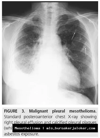
The chest X-ray is often the first imaging study performed when mesothelioma is suspected. While it’s not highly sensitive or specific for mesothelioma, it can reveal abnormalities such as pleural thickening, pleural effusions (fluid accumulation in the pleural space), and masses in the chest. These findings can raise suspicion for mesothelioma and prompt further investigation with more advanced imaging techniques.
The Gold Standard: Computed Tomography (CT)
CT scans, particularly high-resolution CT (HRCT) of the chest, are considered the gold standard for evaluating mesothelioma. CT provides detailed anatomical information, allowing radiologists to visualize the pleura (lining of the lungs), chest wall, mediastinum (the space between the lungs), and abdomen. CT scans can identify pleural thickening, masses, calcifications, and invasion of surrounding structures. Contrast enhancement, achieved by injecting a contrast agent into the bloodstream, helps to differentiate between normal and abnormal tissues.
Magnetic Resonance Imaging (MRI): Superior Soft Tissue Detail
MRI offers superior soft tissue contrast compared to CT, making it particularly useful for evaluating the extent of mesothelioma and its involvement of the chest wall, diaphragm, and mediastinum. MRI can also help to differentiate between mesothelioma and other pleural diseases, such as benign pleural plaques. Specific MRI sequences, such as diffusion-weighted imaging (DWI), can provide information about the cellularity of the tumor and its response to treatment.
Positron Emission Tomography (PET/CT): Metabolic Activity
PET/CT combines the anatomical detail of CT with the functional information of PET. PET uses a radioactive tracer, typically fluorodeoxyglucose (FDG), which is taken up by metabolically active cells, such as cancer cells. PET/CT can help to identify areas of active tumor, assess the extent of disease, and monitor response to treatment. It is particularly useful in distinguishing between benign and malignant pleural thickening and in detecting distant metastases (spread of cancer to other parts of the body).
Advancements in Mesothelioma Radiology by 2025
The field of mesothelioma radiology is rapidly evolving, driven by technological advancements and a growing understanding of the disease. By 2025, we can expect to see significant improvements in imaging techniques, diagnostic accuracy, and personalized approaches to patient care.
Improved Image Resolution and Faster Scanning Times
Advances in CT and MRI technology are leading to higher resolution images and faster scanning times. This allows for more detailed visualization of the tumor and its surrounding structures, with reduced exposure to radiation (in the case of CT) and shorter examination times, improving patient comfort. Techniques like iterative reconstruction in CT and advanced coil technology in MRI contribute to these improvements.
Novel Imaging Agents
Researchers are developing new imaging agents that specifically target mesothelioma cells. These agents can improve the sensitivity and specificity of imaging, allowing for earlier and more accurate detection of the disease. For example, radiolabeled antibodies that bind to specific mesothelioma cell surface markers are being investigated for PET imaging. Such targeted agents could revolutionize mesothelioma diagnosis and treatment monitoring.
Artificial Intelligence (AI) and Machine Learning
AI and machine learning are poised to transform mesothelioma radiology. AI algorithms can be trained to automatically detect and segment mesothelioma tumors on CT and MRI scans, improving diagnostic accuracy and efficiency. AI can also be used to predict treatment response and prognosis based on imaging features. These tools can assist radiologists in making more informed decisions and personalizing patient care.
Radiomics: Extracting Quantitative Data from Images
Radiomics involves extracting a large number of quantitative features from medical images, such as tumor size, shape, texture, and intensity. These features can be analyzed using machine learning techniques to identify patterns that are associated with specific clinical outcomes, such as survival or treatment response. Radiomics holds promise for personalizing mesothelioma treatment and predicting prognosis.
Molecular Imaging: Visualizing Biological Processes
Molecular imaging techniques, such as PET/CT with novel tracers, are allowing researchers to visualize biological processes within mesothelioma tumors, such as angiogenesis (formation of new blood vessels) and immune response. This information can be used to develop and evaluate new therapies that target specific molecular pathways involved in mesothelioma development and progression.
Challenges and Future Directions
Despite the advancements in mesothelioma radiology, several challenges remain. Mesothelioma can be difficult to distinguish from other pleural diseases, such as benign pleural plaques and fibrosis. The disease can also be challenging to stage accurately, particularly in its early stages. Furthermore, there is a need for more effective imaging biomarkers to predict treatment response and prognosis.
Standardization of Imaging Protocols
Standardization of imaging protocols is crucial for ensuring consistent and reliable results across different institutions. This includes standardizing image acquisition parameters, contrast administration protocols, and image interpretation criteria. Standardized protocols will facilitate multi-center clinical trials and improve the comparability of research findings.
Improved Diagnostic Accuracy
Continued research is needed to improve the diagnostic accuracy of mesothelioma imaging. This includes developing new imaging agents, refining imaging techniques, and integrating AI and machine learning into image analysis workflows. Collaboration between radiologists, pathologists, and oncologists is essential for achieving this goal.
Personalized Imaging Strategies
Future research should focus on developing personalized imaging strategies tailored to individual patient characteristics and disease subtypes. This includes identifying imaging biomarkers that predict treatment response and prognosis, and using this information to guide treatment decisions. Personalized imaging strategies have the potential to improve patient outcomes and reduce unnecessary treatments.
Collaboration and Data Sharing
Collaboration and data sharing are essential for accelerating progress in mesothelioma radiology. Sharing of imaging data, clinical data, and research findings will facilitate the development of new diagnostic and therapeutic strategies. Open-source platforms and data repositories can promote collaboration and accelerate the translation of research findings into clinical practice.
Conclusion
Mesothelioma radiology is a dynamic field that is rapidly evolving. By 2025, we can expect to see significant advancements in imaging techniques, diagnostic accuracy, and personalized approaches to patient care. Improved image resolution, novel imaging agents, and the integration of AI and machine learning will revolutionize the diagnosis and management of mesothelioma. Overcoming the remaining challenges through standardization, collaboration, and continued research will be crucial for improving the lives of patients affected by this devastating disease. The future of mesothelioma radiology is bright, with the potential to transform the way we diagnose, treat, and ultimately conquer this challenging cancer.
Conclusion
In conclusion, the role of radiology in the diagnosis, staging, and monitoring of mesothelioma is undeniable. From initial detection through chest X-rays and CT scans to advanced techniques like MRI and PET/CT, radiological imaging provides crucial information for clinicians to effectively manage this aggressive cancer. Understanding the characteristic imaging features of pleural and peritoneal mesothelioma, including pleural thickening, effusions, and tumor spread, is essential for accurate diagnosis and differentiation from other conditions.
As discussed, advancements in imaging modalities continue to improve our ability to detect early-stage disease and assess treatment response. The ongoing refinement of imaging protocols and the integration of artificial intelligence hold tremendous promise for further enhancing the accuracy and efficiency of mesothelioma radiology. We strongly encourage radiologists and other healthcare professionals to stay informed about the latest advancements in this field and to collaborate closely with multidisciplinary teams to ensure optimal patient care. For further information about mesothelioma and available resources, please visit the Mesothelioma.com website.
Frequently Asked Questions (FAQ) about mesothelioma radiology
What are the typical radiological findings on a chest X-ray that might suggest a patient has pleural mesothelioma?
Chest X-rays are often the first imaging study performed when mesothelioma is suspected. While not definitive, certain findings can raise suspicion and warrant further investigation. Common radiological indicators of pleural mesothelioma on chest X-rays include pleural thickening, which can appear as a rind-like density along the lung’s surface. Pleural effusions (fluid accumulation) are also frequently observed. Another sign is volume loss in the affected hemithorax (side of the chest), indicating lung restriction. Sometimes, the mediastinum (the space between the lungs) may shift towards the affected side. It’s crucial to remember that these findings are not specific to mesothelioma and can be caused by other conditions. Therefore, further imaging, such as CT scans or MRI, and ultimately a biopsy, are needed for a definitive diagnosis. While chest X-rays can be a useful initial screening tool, their limitations necessitate more advanced imaging techniques. For more information, you can refer to Mesothelioma as an additional resource.
How does a CT scan help in diagnosing and staging mesothelioma, and what specific features are radiologists looking for?
Computed tomography (CT) scans are crucial for diagnosing and staging mesothelioma. They provide detailed cross-sectional images of the chest and abdomen, allowing radiologists to visualize the extent of the disease. Specifically, radiologists look for circumferential pleural thickening (thickening around the entire lung), nodular pleural masses, and invasion of the mediastinum, chest wall, or diaphragm. CT scans can also identify lymph node involvement, which is important for staging. The presence of pleural effusions, calcifications within the pleura, and encasement of the lung are also important indicators. Furthermore, CT scans can help differentiate between pleural mesothelioma and other conditions that mimic its appearance. Dynamic contrast-enhanced CT scans can also assess tumor vascularity. Based on the CT findings, radiologists can provide a comprehensive assessment of the tumor’s size, location, and spread, which is essential for treatment planning and prognosis.
What role does MRI play in the radiological assessment of mesothelioma, and when is it preferred over a CT scan?
Magnetic Resonance Imaging (MRI) plays a significant role in evaluating mesothelioma, particularly for assessing local disease extent and differentiating it from other conditions. MRI offers superior soft tissue contrast compared to CT scans, making it particularly useful for visualizing the chest wall, diaphragm, and mediastinal structures. This enhanced contrast allows radiologists to better assess tumor invasion into these areas, which is critical for staging and surgical planning. MRI is often preferred over CT when evaluating the diaphragm for tumor involvement or when assessing the chest wall for resectability. It is also useful for differentiating tumor from post-treatment changes like fibrosis. Additionally, MRI avoids ionizing radiation, making it a suitable alternative for patients who require multiple imaging studies. Diffusion-weighted imaging (DWI) during MRI can further characterize the tumor tissue. While CT remains important for overall staging and detecting distant metastases, MRI provides valuable complementary information for local assessment of mesothelioma.